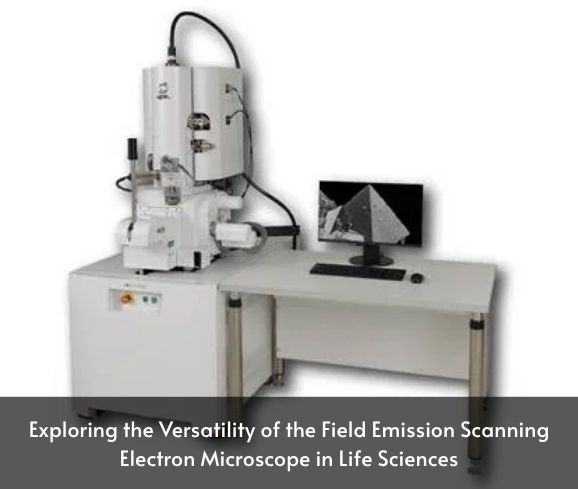
How the Field Emission Scanning Electron Microscope Supports Life Sciences Research
The field emission scanning electron microscope (FE-SEM) has become an indispensable tool in life sciences, offering ultra-high-resolution imaging capabilities that allow researchers to explore biological structures at the nanometer scale. Unlike traditional scanning electron microscopes, FE-SEMs utilize a field emission gun to produce a highly focused electron beam, resulting in superior image resolution and contrast.
In life sciences, FE-SEMs are employed to study a wide range of specimens, including cells, tissues, and biomaterials. Their ability to provide detailed surface morphology and topographical information makes them invaluable for understanding complex biological processes and structures.
Hansvue offers advanced FE-SEM systems tailored for life science applications, ensuring that researchers have access to the latest technology for their investigative needs.
Imaging Cell Structures Using the Field Emission Scanning Electron Microscope
One of the primary applications of the field emission scanning electron microscope in life sciences is the imaging of cellular structures. FE-SEMs provide exceptional surface detail, allowing scientists to observe cell morphology, membrane structures, and intracellular components with remarkable clarity.
This high-resolution imaging is crucial for various studies, including cell differentiation, apoptosis, and interactions between cells and their microenvironment. By visualizing these structures, researchers can gain insights into cellular functions and mechanisms that are essential for advancements in biomedical research.
Hansvue’s FE-SEM systems are equipped with advanced detectors and imaging modes, enabling life science laboratories to capture detailed images of cellular structures efficiently.
Advantages of the Field Emission Scanning Electron Microscope in Microbiology and Histology
In microbiology and histology, the field emission scanning electron microscope offers several advantages over conventional imaging techniques. Its high-resolution capabilities allow for the detailed examination of microorganisms, including bacteria, viruses, and fungi, as well as tissue samples.
FE-SEMs enable the visualization of microbial surfaces, biofilm formations, and interactions between pathogens and host tissues. In histology, they provide detailed images of tissue architecture, aiding in the diagnosis and study of various diseases.
Hansvue’s FE-SEM solutions are designed to meet the specific needs of microbiologists and histologists, offering reliable and high-quality imaging for diverse applications.
Field Emission Scanning Electron Microscope Techniques for Biological Specimen Preparation
Proper specimen preparation is critical for obtaining high-quality images with a field emission scanning electron microscope. Biological samples often require fixation, dehydration, and coating to withstand the vacuum environment and electron beam exposure.
Advancements in preparation techniques, such as cryo-fixation and the use of conductive coatings like gold or platinum, have improved the preservation of biological structures and reduced artifacts. These methods help maintain the integrity of delicate specimens, ensuring accurate imaging results.
Hansvue provides comprehensive support and guidance on specimen preparation protocols, assisting researchers in achieving optimal imaging outcomes with their FE-SEM systems.
Cryo-Imaging with the Field Emission Scanning Electron Microscope in Biomedical Research
Cryo-imaging is a technique that involves rapidly freezing biological samples to preserve their native state for examination under a field emission scanning electron microscope. This method minimizes structural alterations and allows for the observation of specimens close to their natural conditions. en.wikipedia.org
In biomedical research, cryo-FE-SEM is used to study cellular processes, protein interactions, and the ultrastructure of tissues and organs. It provides valuable insights into disease mechanisms and supports the development of new therapeutic strategies.
Hansvue’s FE-SEM systems are compatible with cryo-imaging techniques, offering researchers the tools needed to conduct advanced biomedical investigations.
Field Emission Scanning Electron Microscope Benefits for Tissue Engineering and Biomaterials
Tissue engineering and biomaterials research rely heavily on the detailed analysis of scaffold structures, cell-material interactions, and surface properties. The field emission scanning electron microscope provides high-resolution images that are essential for evaluating the performance and biocompatibility of engineered tissues and materials.
FE-SEMs enable researchers to assess pore sizes, fiber orientations, and the distribution of cells within scaffolds. This information is crucial for optimizing designs and ensuring the success of tissue-engineered constructs.
Hansvue’s FE-SEM offerings are tailored to support tissue engineering applications, providing precise imaging capabilities that facilitate innovation in regenerative medicine.
Hansvue’s Life Science Solutions with the Field Emission Scanning Electron Microscope
Hansvue is committed to advancing life science research by providing state-of-the-art field emission scanning electron microscope systems. Our FE-SEM solutions are designed to meet the diverse needs of biological research, offering features such as:
High-Resolution Imaging: Capturing detailed structures at the nanometer scale.
Advanced Detectors: Enhancing contrast and enabling various imaging.
User-Friendly Interfaces: Simplifying operation and data analysis.
Versatile Sample Compatibility: Accommodating a wide range of biological specimens.
By partnering with Hansvue, life science laboratories gain access to cutting-edge FE-SEM technology and comprehensive support services, ensuring successful research outcomes.
Getting a Field Emission Scanning Electron Microscope for Life Sciences Laboratories
Acquiring a field emission scanning electron microscope for life sciences laboratories involves careful consideration of several factors:
Research Requirements: Determine the specific imaging needs and applications.
Technical Specifications: Evaluate resolution, magnification, and detector options.
Budget Constraints: Assess the total cost of ownership, including maintenance and training.
Vendor Support: Ensure the availability of technical assistance and service agreements.
Hansvue offers a range of FE-SEM systems tailored for life science applications, along with expert consultation to help laboratories select the most suitable equipment for their research goals.
Conclusion
The field emission scanning electron microscope has revolutionized life sciences by providing unparalleled imaging capabilities that reveal the intricate details of biological specimens. Hansvue’s commitment to delivering advanced FE-SEM solutions empowers researchers to explore new frontiers in biology, medicine, and biotechnology.